Image Gallery : Photo Gallery
HOME > Image Gallery > Photo Gallery
Kaolin Minerals
Please click to view a larger image.
1. SEM image of kaolinite
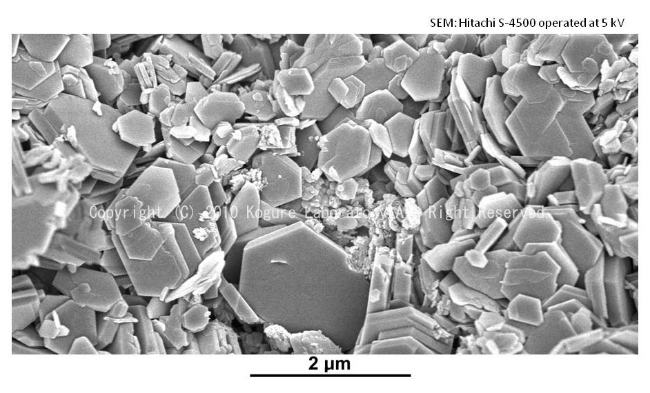
SEM image of kaolinite. Kanpaku mine, Tochigi, Japan (hydrothermal origin).
![]()
2. SEM image of kaolinite with a vermicular form
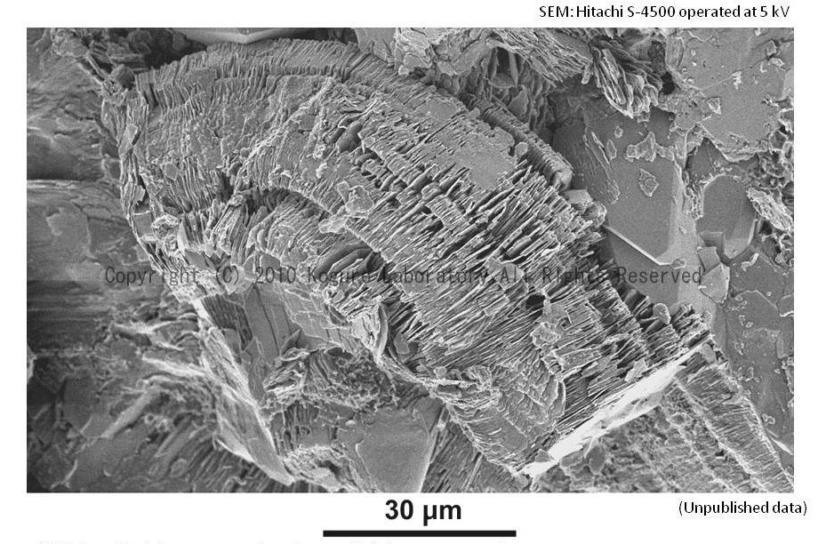
SEM image of kaolinite with a vermicular form, found in the sandstone reservoir, the North Sea.
Kameda, J., K. Saruwatari, D. Beaufort and T. Kogure: "Textures and polytypes in vermiform kaolins diagenetically formed in a sandstone reservoir: a FIB-TEM investigation", Euro. J. Mineral., 20 (2008) 199-204.
![]()
3. SEM images of halloysite grown radially
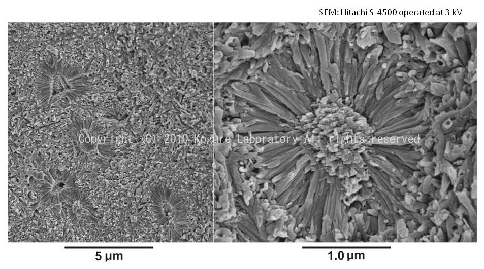
SEM images of halloysite grown radially, from Eureka, Nevada, USA (S.W. Bailey collection). The material at the center of the halloysite spokes is gibbsite.
![]()
4. EBSD and Kikuchi patterns from kaolin group minerals
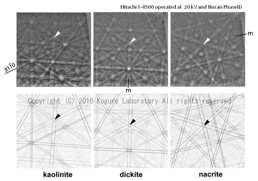
EBSD patterns from the basal (001) plane of kaolin group minerals (kaolinite, dickite, nacrite) and corresponding drawings of the Kikuchi patterns.
Kogure, T., A. Inoue and D. Beaufort: "Polytype and morphology analyses of kaolin minerals by electron back-scattered diffraction", Clays Clay Miner., 53 (2005) 201-210.
![]()
5. Simulated HRTEM images of kaolin and corresponding structure models
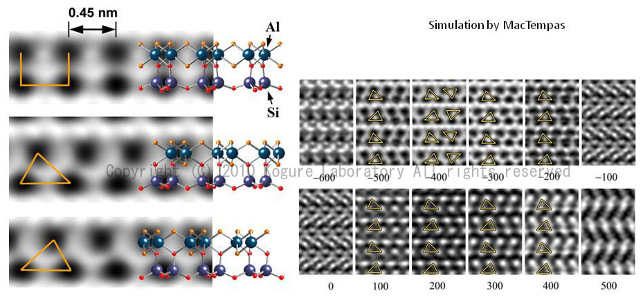
(Left) Simulated HRTEM images of kaolin 1:1 layer observed along Xi (i = 1,2,3) directions and corresponding structure models. Vacc 200 kV, Cs 0.5 mm, Spread of focus 10 nm, beam convergence 0.5 mrad, defocus -40 nm, and specimen thickness 2.5 nm were used for the simulation. (Right) Defocus (in Å) dependence of the simulated contrast of dickite observed along [100].
Kogure, T. and A. Inoue: "Stacking defects and long-period polytypes in kaolin minerals from a hydrothermal deposit", Eur. J. Mineral., 17 (2005) 465-474.
![]()
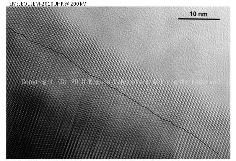
6. HRTEM image of dickite from the sandstone oil reservoir of the North Sea
HRTEM image of dickite recorded along [100]. The specimen was obtained from the sandstone oil reservoir of the North Sea. The crystal was partially damaged by beam radiation. The image shows two-layer periodicity with no stacking disorder.
Kogure, T. and A. Inoue, Determination of defect structures in kaolin minerals by High-Resolution Transmission Electron Microscopy (HRTEM) , Am. Mineral., 90 (2005) 85-89.
![]()
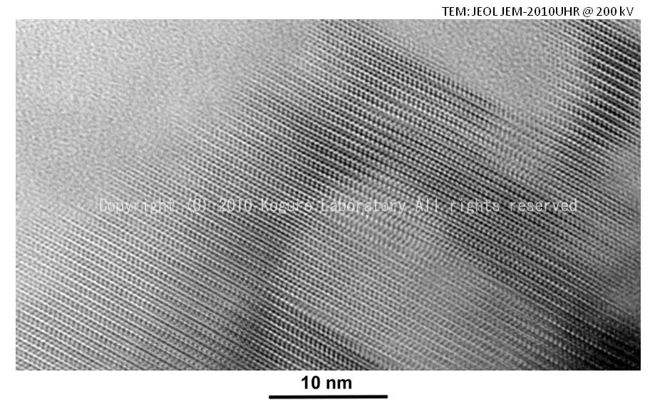
7. HRTEM image of kaolinite from the sandstone oil reservoir of the North Sea
HRTEM image of kaolinite from the sandstone oil reservoir of the North Sea, showing dense stacking disorder. The solid line indicates the stacking disorder which is formed by alternative interlayer displacements between the adjacent layers. Disorder of the octahedral vacancy site in the dioctahedral sheet is not obsreved in this specimen.
Kogure, T. and A. Inoue, Determination of defect structures in kaolin minerals by High-Resolution Transmission Electron Microscopy (HRTEM) , Am. Mineral., 90 (2005) 85-89.
![]()
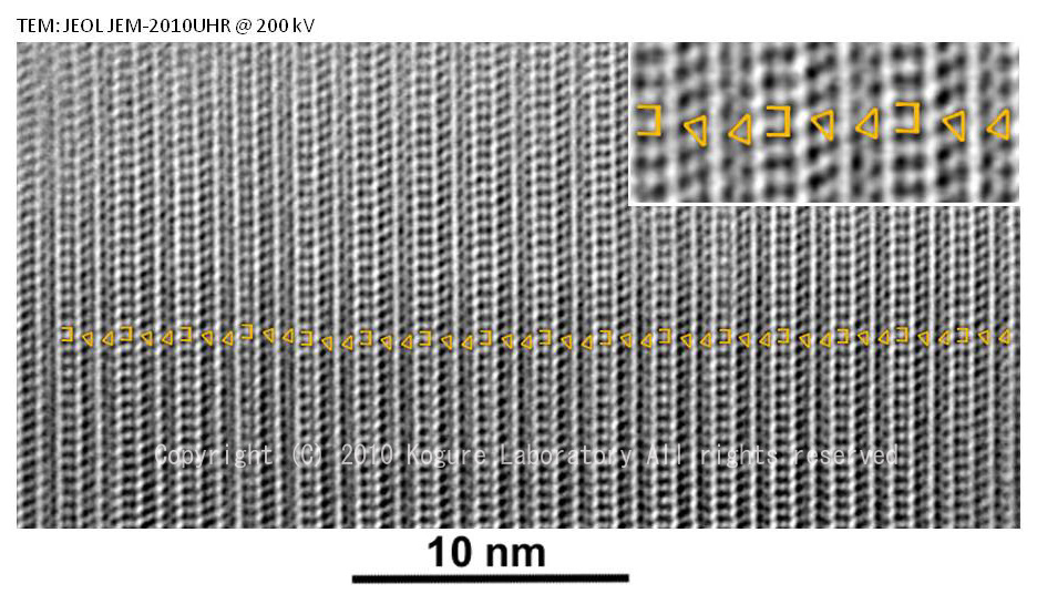
8. HRTEM image of the kaolin specimen of hydrothermal origin -1
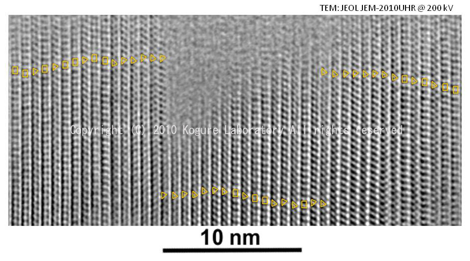
HRTEM image of the kaolin specimen of hydrothermal origin (Kasuga mine, Kagoshima, Japan). The triangular and rectangular patterns on the image indicates the contrast in each 1:1 layer. The disorder of the patterns corresponds to that of the octahedral vacancy site. This crystal also contains disorder of the interlayer displacement, resulting in completely random stacking.
Kogure, T. and A. Inoue: "Stacking defects and long-period polytypes in kaolin minerals from a hydrothermal deposit", Eur. J. Mineral., 17 (2005) 465-474.
![]()
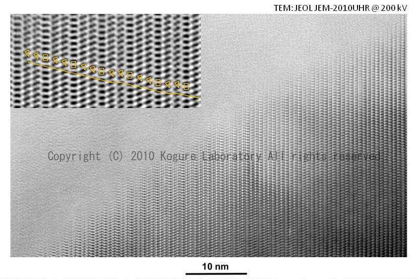
9. HRTEM image of the kaolin specimen of hydrothermal origin -2
HRTEM image of the kaolin specimen of hydrothermal origin (Kasuga mine, Kagoshima, Japan), with a three-layer stacking structure consisting of three alternating layers with different octahedral vacancy sites.
Kogure, T. and A. Inoue: "Stacking defects and long-period polytypes in kaolin minerals from a hydrothermal deposit", Eur. J. Mineral., 17 (2005) 465-474.
![]()
10. HRTEM image of the kaolin specimen of hydrothermal origin -3
HRTEM image of the kaolin specimen of hydrothermal origin (Kasuga mine, Kagoshima, Japan), showing a different three-layer stacking from that in the previous slide. It is likely that such stacking structure was generated by the spiral growth mechanism.
Kogure, T. and A. Inoue: "Stacking defects and long-period polytypes in kaolin minerals from a hydrothermal deposit", Eur. J. Mineral., 17 (2005) 465-474.
![]()
11. HRTEM images of the kaolinite specimen of sedimentary origin
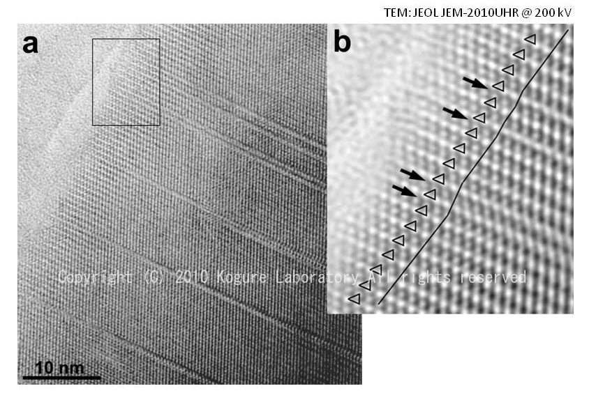
(Left) HRTEM images of the kaolinite specimen of sedimentary origin (Brazil, Capim), showing linear contrasts originating from stacking faults. (Right) More magnified image around the specimen edge , indicating that the faults are due to the different interlayer shift.
Kogure, T., C.T. Johnston, J.E. Kogel and D. Bish: "Stacking disorder in a sedimentary kaolinite", Clays Clay Miner., 58 (2010) 63-72.
![]()