Image Gallery : Photo Gallery
HOME > Image Gallery > Photo Gallery
Biominerals
Please click to view a larger image.
1. Polymorphs of CaCO3 (SEM)

Polymorphs are crystalline materials with the same chemical composition but different crystal structures. In the atomosphic pressure, calcium carbonate (CaCO3) has three polymorphs: a) calcite, b) aragonite, and c) vaterite. These pictures show the morphologies of the three polymorphs, precipitated from the CaCO3 supersaturated solution. Calcite frequently shows a rhombohedron surrounded with smooth surfaces. Aragonite generally adopts an acicular shape with a hexagonal cross-section. Vaterite often shows plate or disc like appearance. However, biotic CaCO3 crystals often have completely different morphologies from those with abiotic processes.
![]()
2. Amorphous calcium carbonate (ACC) in gastrolith of a fresh-water crayfish (SEM)
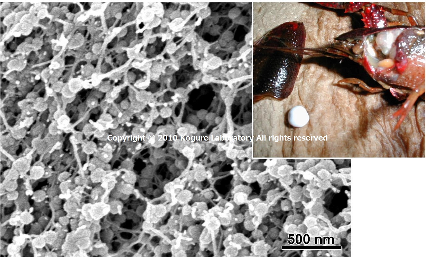
Although most of biotic calcium carbonate are crystalline, a few adopts an amorphous form which is called amorphous calcium carbonate (ACC). A fresh-water crayfish, Procambarus clarkia, stores ACC in its gastrolith for molting. ACC in the gastrolith forms as spherules, as shown in this image. Such ACC with a large surface area easily dissolve under specific solution condition and the crayfish can supply calcium from the gastrolith to make hard exoskeleton.
![]()
3. Asteriscus of salmon, Oncorhynchus keta(SEM)
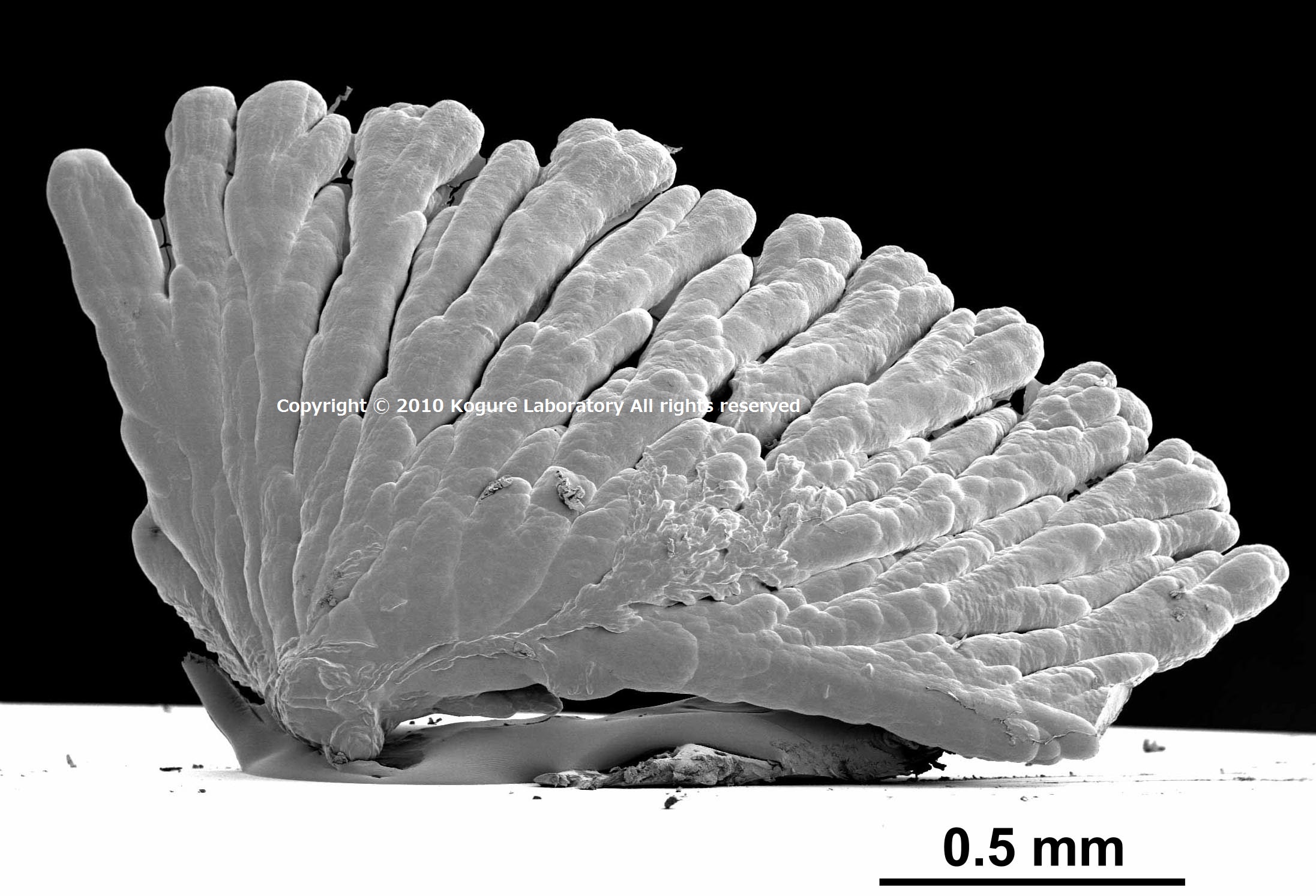
Among the polymorphs of calcium carbonate, vaterite is less common than calcite and aragonite. Asteriscus, one of the fish otoliths, is a rare example of the biominerals formed with vaterite. This picture is a low magnification SEM image of asteriscus of salmon, Oncorhynchus keta, showing a distinct morphology, like micron-sized coral.
![]()
4. Nacreous layers of mollusk shells(SEM)

Inside of some mollusk shells shows beautiful iridescence and is used as jewelry. This iridescence originates from platy crystals of aragonite intercalated with thin organic films. This structure is called the nacreous layer or just nacre. The left picture is the nacreous layer of a bivalve Pinctada fucata, and the right one is that of a gastropod, Omphalius rusticus. The structure in the right is expressed as “stack of coins” figuratively.
![]()
5. Surface of the prismatic layer of Pinctada fucata etched by EDTA
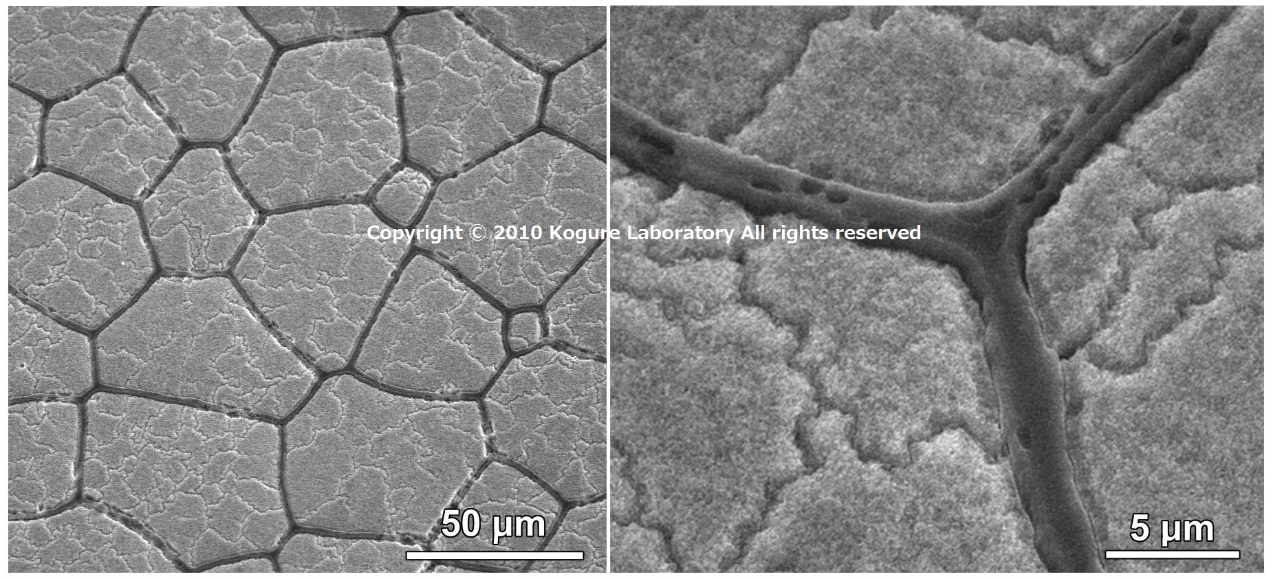
The Outside of the pearl oyster shell forms the prismatic layer which consists of the calcite prisms. These pictures show the surface of the layer after etching by EDTA (left: lower magnification, right: higher magnification). The dark colored interprismatic frames are made of organic matrix. If the calcite prism is etched by EDTA, sinuous lines appear on the surface. They are grain boundaries inside the prism.
![]()
6. Crossed lamellar structure in a limpet shell(SEM)

This picture shows the inner surface of the crossed lamellar layer in a limpet shell (Lottia kogamogai). This shell has five layers laminated along the thickness direction, and the first and third layer adopt the crossed lamellar structure. The structure is built with aragonite flat prisms. The prisms are stacked obliquely against the surface and this oblique direction changes alternately, as shown in this figure.
![]()
7. Spine of purple sea urchin (SEM)
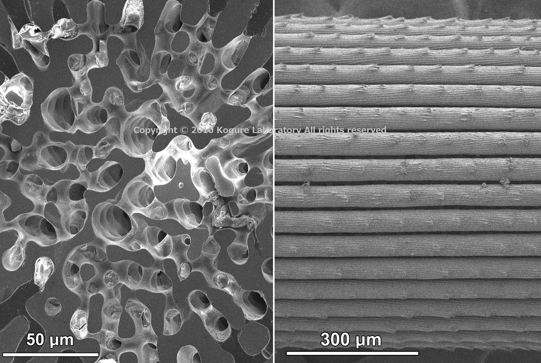
The spine of sea urchins consists of calcite containing a considerable amount of magnesium. In spite of its complicated morphology, the spine is a single crystal of calcite with its c-axis parallel to the spine. However, its mechanical properties are completely different from abiotic single-crystalline calcite. The left of the figure indicates the central part of the cross-section of the spine from purple sea urchin, Anthocidaris crassispina and the right one is a part of the longitudinal surface of the spine, after bleaching with NaClO.
![]()
8. Coccolith (SEM)

Coccoliths are calcified scaled formed around the cell of coccolithophores, marine phytoplankton. This picture shows a coccolith of Pleurochrysis carterae (top left: the whole picture of the coccolithophore). The coccolith ring consists of about twenty calcite crystal units, with two types of crystals allocated alternately along the circumference. The two types have different shapes and crystal orientations. The regulation mechanism of the coccolith structure is one of the most interesting topics of biomineralization.
![]()
9. High-resolution structure image of aragonite (TEM)

After the appearance of the first TEM in 1932, the continued improvements of TEM technology has enabled to observe directly the atomic arrangements in crystals. This picture is a TEM image of the aragonite crystal in the nacreous layer of a bivalve Pinctada fucata, with the beam direction parallel to [100] of aragonite. The repeated dark contrasts indicated with the red arrows in the figure correspond to the circled atomic groups in the structure model of aragonite at the top left.
![]()
10. Larval shell of bivalve, Pinctada fucata (TEM)
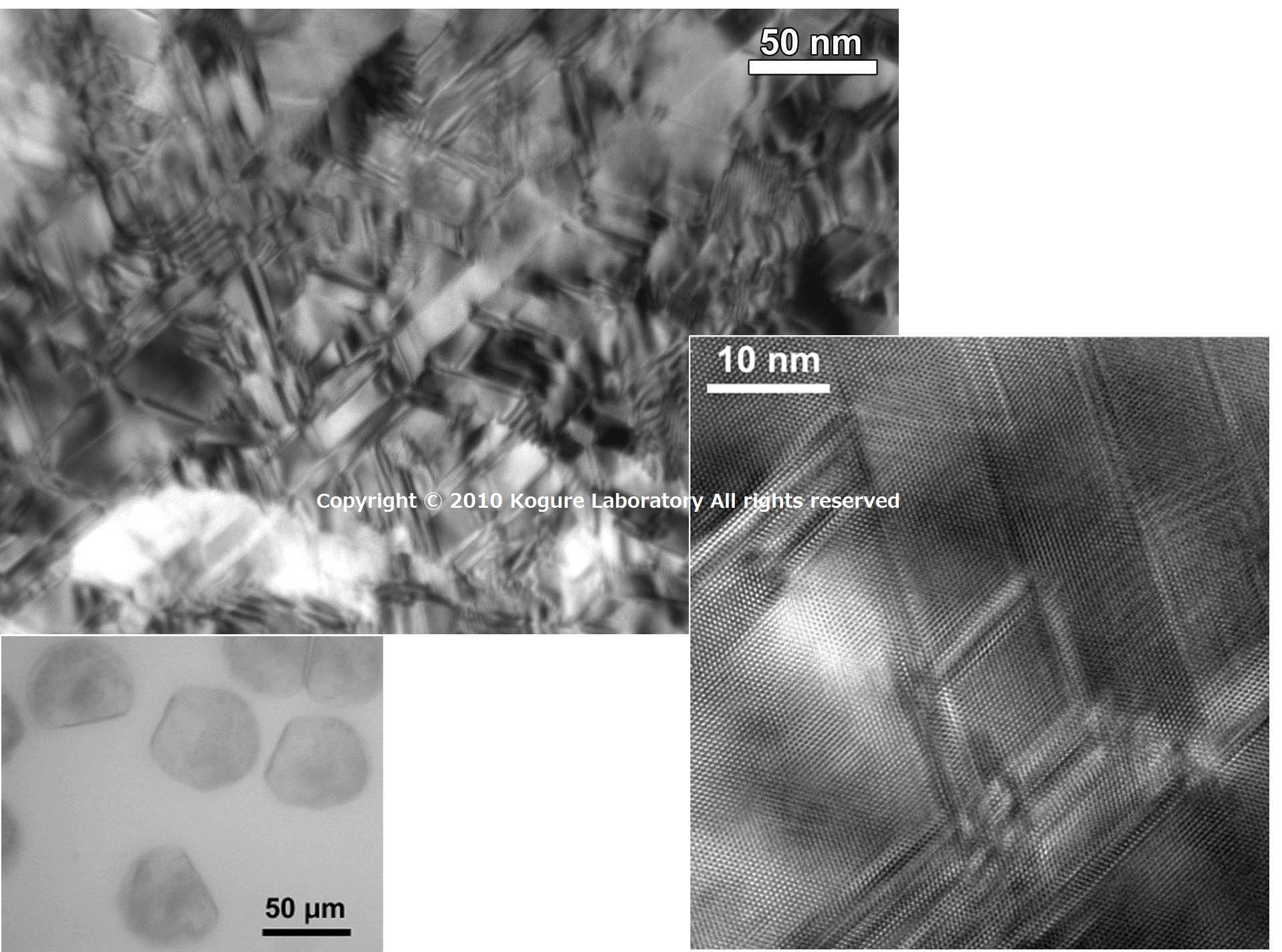
Over a dozen hours after fertilization, the embryo of bivalves is surrounded by larval shell (bottom-left). This TEM micrograph is the plan-view bright-field image of the larval shell of a pearl oyster, Pinctada fucutta. The complex contrast is corresponding to the multiple {110} twins in the aragonite crystal, which is more distinct in the high-resolution TEM image at the bottom-right.
![]()
11. Ligament of bivalve, Pinctada fucata ligament (TEM)
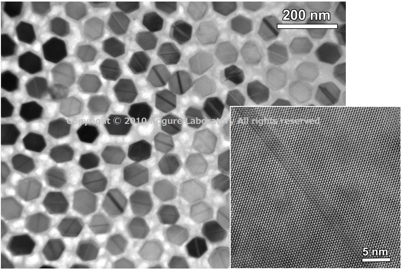
The ligament of bivalves has the role of hinge and consists of the bundles of acicular aragonite crystals. This TEM picture shows the ligaments of Pinctada fucuta, where the hexagonal crystals are the cross-section of the acicular aragonite buried in organic matter. A band contrast along the diagonal of the hexagon is the {110} twin of aragonite, as shown in the high-resolution TEM image at the bottom-right.
![]()
12. Magnetosome in magnetotactic bacteria (TEM)
Magnetosome is a fine particle of magnetite (Fe3O4), formed inside of the cell by magnetotactic bacteria. This is a kind of magnetism sensor with which the bacteria finds their way in water. One magnetosome is tens of nanometers in size. This TEM image clearly shows the formation of multiple {111} twins inside the particle.
![]()