Research Subjects and Facilities
HOME > About Us
Kogure Laboratory is
Kogure laboratory belongs to the Department of Earth and Planetary Sciences, Graduate School of Science, The University of Tokyo.
The main target of the research in the laboratory is to elucidate the structures of natural inorganic materials formed around the surface of the earth (biosphere) and their formation processes. Electron microscopy (both TEM and SEM) is primarily applied in the research, which is another characteristic of the laboratory. At present we forward the research, especially focusing on the following subjects.
Research Subjects
Formation mechanism of biominerals, inorganic materials made by organisms
Living organisms often utilize inorganic materials (biominerals) as their hard tissues like bone, teeth, exoskeleton, shell, scale, etc. It is well known that our bone and teeth are made of calcium phosphates (e.g., hydroxyapatite) with organic matrices. On the other hand, shell of mollusks, exoskeleton of crustacean, eggshell, and otolith are made of calcium carbonate. These hard tissues often exhibit excellent properties to sustain the activity of the organisms, which are generally originated from well regulated micro- (or nano-) structures (crystalline phase, size distribution, morphology, defect, crystal orientation) of the biominerals in the tissues. Another characteristic of the biominerals is that they are formed in environments where organisms live, i.e. at normal temperature and pressure, and relatively in a short time, which is largely different from minerals in rocks. This means that kinetics often play an important role with thermodynamics. The formation and regulation mechanisms of biominerals have been investigated for a long time, but still not well understood.
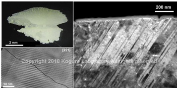 (Top left) Optical micrograph of otolith in salmon.
(Right) Dark-field TEM image of the otolith to show high density of {110} aragonite twins.
(Bottom-left) HRTEM image of the twins.
It is known that most biominerals nucleate on organic matrix which may regulate the crystal orientation and possibly crystalline phase, and they grow in solution incorporating organic molecules secreted from the organism. These intracrystalline organic molecules may also influence the structure of biominerals. We are investigating the roles of these organic matrix and intracrystalline molecules in the formation processes of biominerals, by analyzing fine structures in the hard tissues mainly using TEM/SEM, and/or by conducting in vitro experiments of crystal growth. The research of biominerals is definitely an interdisciplinary science. We often collaborate with other laboratories with different methodologies to advance the research.
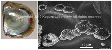 SEM image of the initial calcified structure for the nacreous layer of a pearl oyster, Pinctada fucata (left), formed on the organic sheet which covers the preexisting prismatic outer layer.
(Saruwatari et al., 2009)
- Representative recent publications
- ☻Suzuki, M., J. Kameda, T. Sasaki, K. Saruwatari, H. Nagasawa and T. Kogure: "Characterization of the multilayered shell of a limpet, Lottia kogamogai (Mollusca: Patellogastropoda), using SEM–EBSD and FIB–TEM techniques", J. Struct. Biol., 171 (2010) 223-230. DOI: 10.1016/j.jsb.2010.04.008
- ☻Kudo, M., J. Kameda, K. Saruwatari, N. Ozaki, K. Okano, H. Nagasawa and T. Kogure: " Microtexture of larval shell of oyster, Crassostrea nippona: a FIB-TEM study", J. Struct. Biol., 169 (2010) 1-5. DOI: 10.1016/j.jsb.2009.07.014
- ☻Suzuki, M., K. Saruwatari, T. Kogure, Y. Yamamoto, T. Nishimura, T. Kato and H. Nagasawa: "An acidic matrix protein, Pif, is a key macromolecule for nacre formation", Science, 325 (2009) 1388-1390. DOI: 10.1126/science.1173793
- ☻Saruwatari, K., T. Matsui, H. Mukai, H. Nagasawa and T. Kogure: "Nucleation and growth of aragonite crystals at the growth front of nacres in pearl oyster, Pinctada fucata", Biomaterials, 30 (2009) 3028-3034. DOI: 10.1016/j.biomaterials.2009.03.011
Fine structures of clay minerals and related layered materials
If the minerals formed at a high temperature and/or pressure deeply under the ground are exposed to surface environments, they decompose and transform to new minerals, which are often called “secondary minerals”. Clay minerals are representative secondary minerals abundant on the earth or in biosphere. Clay minerals form as very fine particles which we cannot recognize visually or even if using optical microscopy. However, they are very important and intimate materials for our life. For instance, clays are main inorganic constituents of soil, where they retain water, supply nutrients, or adsorb toxic elements. Clays are the raw materials of ceramic industries, and furthermore, can be new materials for future, sustainable society.
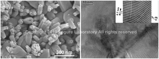 (Left) SEM image of halloysite tubes formed from weathering of feldspar.
(Right) HRTEM image of the cross-section of a chrysotile tube (polygonal serpentine).
Many clay minerals belong to phyllosilicates (or sheet silicates). The structures of phyllosilicates are characterized by infinite sheets made of SiO4 tetrahedrons. Moreover, the tetrahedral sheets combine with octahedral sheets, alkaline metals, water molecules, etc. to form a layer as a structure unit of phyllosilicates. The internal structure of the unit layer is rather rigid, but mutual position between the adjacent layers is often variable, these variables result in the formation of "polytypes", a special case of polymorphs for layered materials, and/or dense stacking faults.
As mentioned above, clay minerals are so fine and their stacking structures can be varied that their true structures have not been well understood yet. Traditionally powder X-ray diffraction, thermal analyses, infrared spectroscopy, etc. are applied to identify or investigate the structures of clays. However, such “macroscopic” analyses often do not give solutions for the understanding of the clay structures. On the other hand, high-resolution transmission electron microscopy (HRTEM) is a powerful tool to determine the clay structures by observing directly their atomic structures, from which we can readily understand polytypes, stacking faults, interstratification in clays. However, HRTEM of clays has several obstacles. Radiation damage by electron beam is the most serious one. We are challenging to reveal the true structures of clays, by applying state-of-the-art electron microscopy to them and overcoming these obstacles.
 (Left) Crystal structure of trioctahedral mica
(Right) HRTEM image of kaolinite with enantiomeric twins domains
- Representative recent publications
-
- ☻Kogure, T., C.T. Johnston, J.E. Kogel and D. Bish: "Stacking disorder in a sedimentary kaolinite", Clays Clay Miner., 58 (2010) 63-72. DOI: 10.1346/CCMN.2010.0580106
- ☻Kameda, J., K. Saruwatari, D. Beaufort and T. Kogure: "Textures and polytypes in vermiform kaolins diagenetically formed in a sandstone reservoir: a FIB-TEM investigation", Euro. J. Mineral., 20 (2008) 199-204. DOI: 10.1127/0935-1221/2008/0020-1806
- ☻Kogure, T., J. Kameda and V.A. Drits : "Stacking faults with 180°layer rotation in celadonite, iron and magnesium-rich dioctahedral mica", Clays Clay Miner., 56 (2008) 612-621. DOI: 10.1346/CCMN.2008.0560602
- ☻Kogure, T. and J. Kameda: " High-resolution TEM and XRD simulation of stacking disorder in 2:1 phyllosilicates", Zeit. Kristallogr., 223 (2008) 69-75. DOI: 10.1524/zkri.2008.0004
- ☻Kogure, T., J. Kameda and V.A. Drits: "Novel 2:1 structure of phyllosilicates formed by annealing Fe3+, Mg-rich dioctahedral mica", Am. Mineral., 92 (2007) 1531-1534. DOI: 10.2138/am.2007.2667
Developments of new techniques using electron beam to investigate minerals
Most minerals are crystalline. Crystals are characterized with the repeated units in their atomic structures, termed “unit cell”. After the discovery of X-ray and subsequent establishment of diffraction theory, people devoted to determine the atomic arrangement in the unit cell in minerals, mainly using X-ray diffraction. Owing to their enormous efforts, the unit cell structures of most minerals have been precisely determined in the last century, which is the fundamental knowledge to understand the properties of minerals. However, the atomic arrangement in the unit cell represents only the “average” structure of the whole crystal and is not enough to explain various phenomena in mineralogy. For instance, morphology of minerals is primarily determined by the surface energy of crystals, and the surface energy depends on the atomic arrangement at the surfaces to terminate three-dimensional crystal structures. However, the surface atomic structures of minerals are generally not well understood yet. Including such surface structures, understanding of local structures in minerals is necessary for the advance of mineralogy and terrestrial material science. For this purpose, new ideas, methods, or techniques must be introduced or developed in this science field, which another subject of our laboratory.
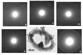 Application of orientation analyses using Kikuchi-patterns generated by a convergent beam, to biominerals
(Saruwatari et al., 2008)
- Representative recent publications
- ☻Kogure, T. and E. Okunishi: "Cs-corrected HAADF-STEM imaging of silicate minerals", J. Electron Microsc., 60 (2010) 1-9. DOI: 10.1093/jmicro/dfq003
- ☻Rozhdestvenskaya, I. V., T. Kogure, E. Abe and V.A. Drits: "A structural model for charoite", Miner. Mag., 73 (2009) 883-890. DOI: 10.1180/minmag.2009.073.2.883
- ☻Saruwatari, K., J. Akai, Y. Fukumori, N. Ozaki, H. Nagasawa and T. Kogure: "Crystal orientation analyses of biominerals using Kikuchi patterns in TEM", J. Mineral. Petrol. Sci., 103 (2008) 16-22. DOI: 10.2465/jmps.070611
- ☻Kameda, J., R. Inoguchi, D.J. Prior and T. Kogure: “Morphological analyses of minute crystals by using stereo-photogrammetric scanning electron microscopy and electron back-scattered diffraction", J. Microscopy, 228 (2007) 358-365.
- ☻Kudoh, Y., J. Kameda and T. Kogure: "Dissolution of brucite on the (001) surface around neutral pH: in-situ AFM observation", Clays Clay Miner., 54 (2006) 598-604. DOI: 10.1346/CCMN.2006.0540506
![]()
Facilities
200kV High-Resolution Transmission Electron Microscope: TEM (HEOL JEM-2010)
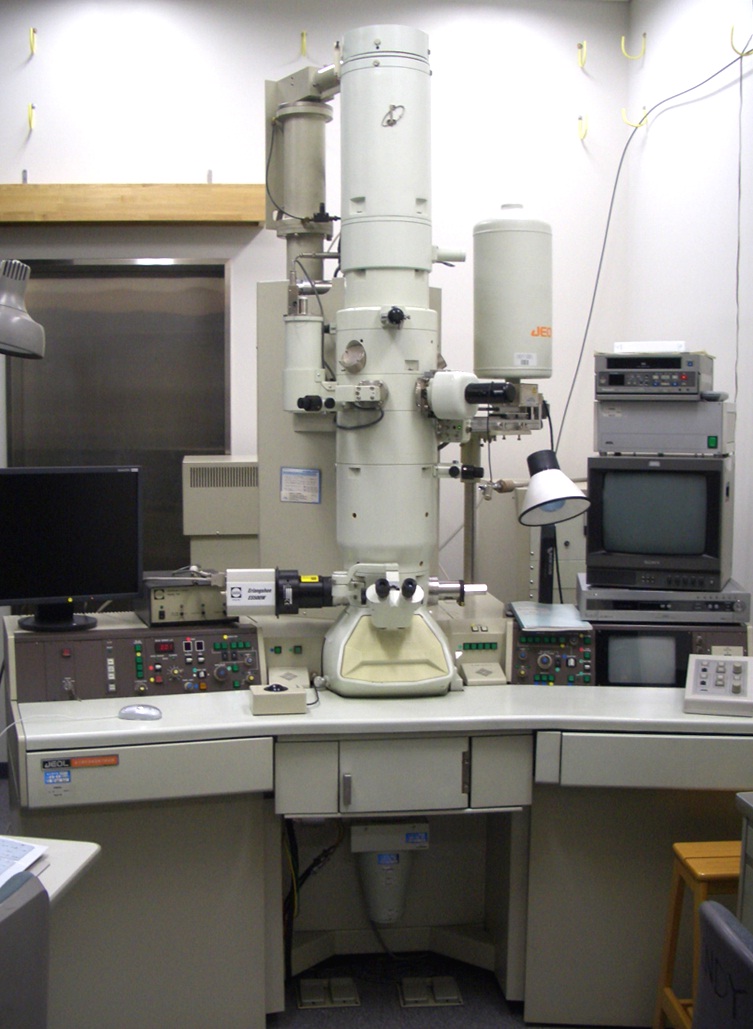
Year Equipped: 1993
This is a 200kV transmission electron microscope especially for high-resolution imaging. A point resolution of about 2.0Å is attained owing to a low aberration objective lens, which enables direct visualization of atomic arrangements in minerals. An energy dispersive X-ray detector and two types of CCD cameras are equipped to the TEM.
Cold-type Field-Emission Scanning Electron Microscope: Cold-FE SEM (Hitachi S-4500)
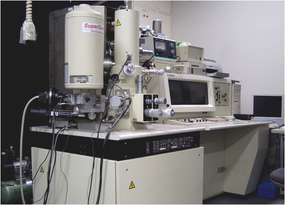
Year Equipped: 1996
This scanning electron microscope (SEM) has a cold-type field-emission electron gun which enables high-resolution imaging owing to its high brightness and small energy spread. This SEM also possesses six kinds of signal detectors around the specimen chamber to acquire various information from the specimen.
Focused Ion Beam System: FIB (Hitachi FB-2100)
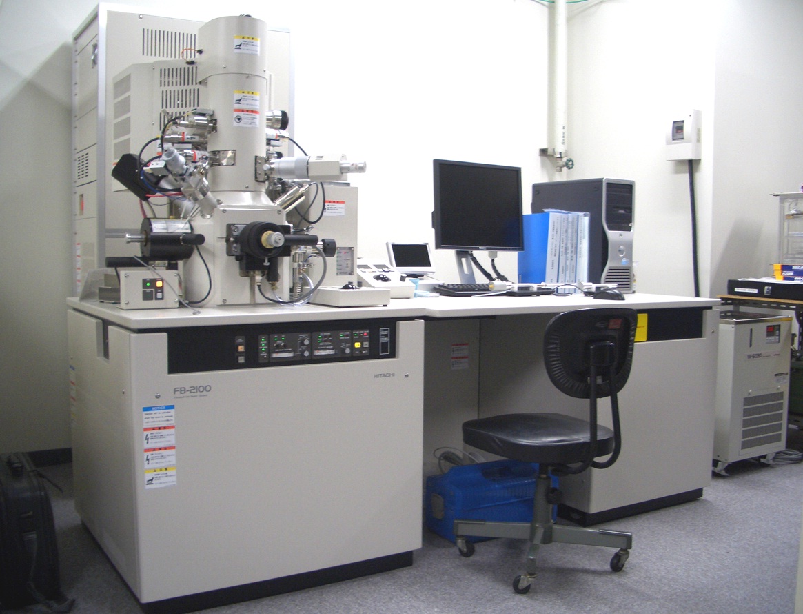
Year Equipped: 2006
This apparatus is used for the preparation of TEM specimen from specific points on bulk specimens or microprocessing, using a focused ion beam of gallium. Especially the micro-sampling system is equipped to make TEM specimens efficiently.
Left: TG-DTA (Rigaku Thermo plus EVO TG8120)
Right: Powder XRD (Rigaku RINT Ultima+) (Common use)
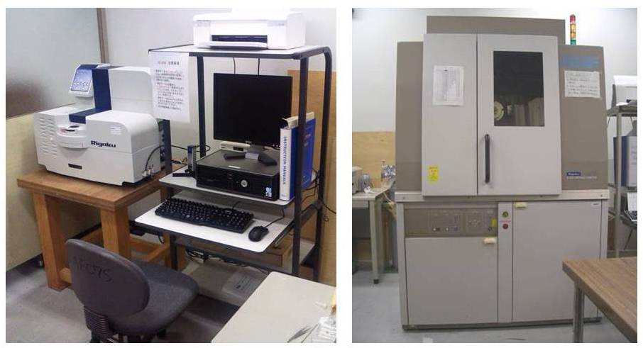
<TG-DTA> Year Equipped: 2009
The highest temperature: 1500°C. The machine is used to determine clay phases and quantification of the organic content in biominerals.
![]()